
The Microscope
Skills
1. Use a light microscope to investigate the structure of cells and tissues, with drawing of cells
2. Drawing of cell structures as seen with the light microscope. Demonstrate how to draw cell structures seen with a microscope using sharp, carefully drawn lines and straight edge lines for labels.
Objectives
- Label the parts of the microscope
- Define magnification
- Given the magnification of the ocular and objective lens, calculate the total microscope magnification.
- Measure the field of view diameter of a microscope under low. medium and high power.
- Estimate the size of the sample in the microscope field of view
- Demonstrate how to focus on a sample
- Demonstrate how to draw cell structures seen in a microscope using the rules for Biological Drawings.
- Ensure you have a completed a labelled microscope drawing.
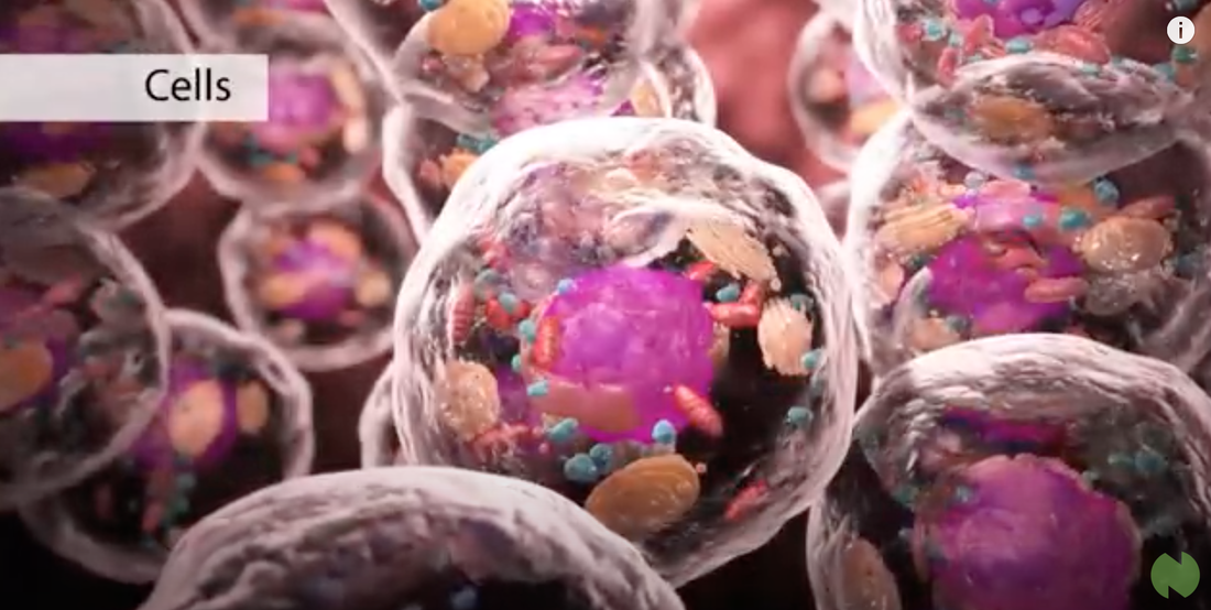
- You have chosen 3 of the 5 Cell images from Cells Online.
- Ensure the Virtual lab and worksheet is completed.
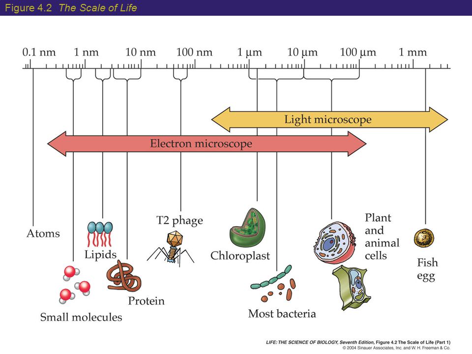
- Make notes on cell size.
- Enjoy the you tube clips. click the image - it is linked to you tube,
- LEARN THE SONG :) HAVE FUN - Maxine.
- Tomorrow is a lesson on Prokaryotes and Eukaryotes

Parts of the Compound Microscope
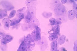
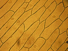
Exercise: Choose three types of cells to draw - USE: Rules for Biological Drawing and 3 images from Cells online -see below
|
Use of the Compound MicroscopeComplete the Introduction to the Virtual Microscope and its Parts
Exercise: Choose a PPTX to learn a little more about microscopes and cells. Use some of the information creatively in your ML book.
|
Cell SizePress the button below which will take you to an interactive task on cell size
| ||||||||||||||||||||||||||||


|
|
